

Digital Panorex
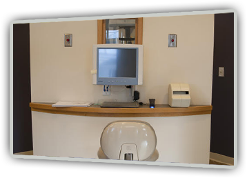 Our office uses digital technology for Panorex images that are clear, accurate, and easily stored and transferred. Best of all, a digital Panorex can be taken with up to 90% less radiation than a traditional Panorex. We can review the digital Panorex images with you from our chairside monitor, explaining what the dentist sees and helping you make an informed decision
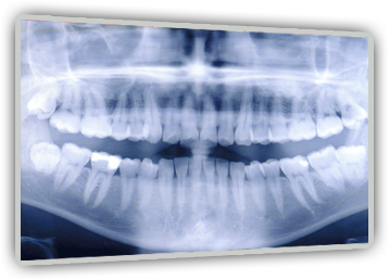
A Panorex image is an X-ray that depicts a panoramic view of your mouth, from one side to the other. This image can help the dentist understand the relationship between your teeth, jaws, and occlusion (bite, or how your top and bottom teeth fit together). A panorex imagine is often used as a screening tool for diagnosing tempormandibular joint disorders, wisdom teeth development, congentially missing permanent teeth, and detecting lesions within the jaw bone. When planning extensive restorative or cosmetic cases, a Panorex may be necessary in addition to intra- and extra-oral photos and digital X-rays.
Digital Radiography
What We See Is What We Get
We've invested in a new way of looking into your mouth – a procedure that's fast, comfortable, and incredibly precise. Using digital radiography, we can clearly identify all external and internal anatomical structures and accurately diagnose your dental problems. Even more amazing, we can immediately translate that information into a large, clear, accurate image, projected onto a monitor that patient and doctor can study together in the operatory. You won’t even have to leave your chair. Digital radiography’s technology improves and simplifies the way we care for our patients’ teeth, resulting in better dental evaluations and treatment decisions. As the most important member of your dental team, you need to understand the condition of your mouth, as well as our recommendations for treatment. Digital radiographs help us help you.
Reduced Radiation, Radical Results
Traditionally, dentists used X-rays to see what the naked eye could not; X-rays were developed in a darkroom with hazardous chemicals, and then viewed on a special light board. The developed X-rays had to be stored, which required large filing systems. By far, the worst part of traditional X-rays was the radiation exposure to patients. Digital radiography has completely transformed this process.
Now, when you come into the office for X-rays, a tiny sensor is placed in your mouth to emit a small amount of radiation – up to 90-percent less than traditional X-rays required. This creates a detailed image of your internal oral structures that is immediately viewable on a chairside monitor, carrying with it all the conveniences of other digitized images. We can rotate and magnify it, adjust it for contrast, and even color-code it for educational purposes. The digital images store easily and efficiently in our computer files, safe and sound. For insurance purposes, referrals, or patient education, digital X-rays can be easily, inexpensively, and accurately reproduced indefinitely.
Digital X-rays offer unparalleled benefits over traditional radiographs: they’re convenient, safe for the environment, provide a great opportunity for patient education, can be transferred and copied accurately, and best of all, they’re safer for our patients.
We invite you to call Commencement Bay Dentistry of Tacoma, WA today to schedule your appointment with Dr. Miller. Our convenient location is accessible to patients from Tacoma and the surrounding areas. We look forward to learning how we can make you smile!